The goals of this research
are (1) to determine the growth and motion of contact regions and the
associated force variations over time between the human fingerpad and
carefully chosen transparent test objects whose microtexture, shape or
softness is varied in a controlled manner and (2) Experimental measurement
of the surface deformations of human fingertips under shaped indentors.
The results obtained are being used to gain a deeper
understanding of the neurophysiological and psychophysical data we
have already obtained for the same test objects.
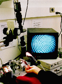
To measure the in vivo surface deformations of the fingerpad under various
tactile stimuli, we have designed a videomicroscopy system together with
a high precision tactile stimulator. The videomicroscopy system consists
of a set of video zoom lenses attached to a high-resolution CCD camera,
whose output can either be digitized into the computer system memory in
real time at about 20 frames/s, or stored on a laserdisk at 30 frames/s
for off-line digitization. The zoom lenses enable continuous variation
of magnification, with the field of view covering the entire fingerpad,
or just a few fingerprint ridges. The tactile stimulator is composed of
a linear stepper motor with a microstepping drive. The motor is controlled
by a 80386 PC, with a specified indentation velocity commanded by a 80486
PC via a digital link. To record the contact force, a strain gage based
single degree of freedom force sensor is mounted on the motor to which
a transparent test object can be attached for both biomechanical and psychophysical
experiments. This method allows the force and video data to be synchornized
and stored in the 80486 PC. With this setup, we are able to investigate
how the skin-object contact region changes with indentation velocity and
force. In active touch experiments the subject contacts a stationary specimen,
whereas in passive touch experiments the stimulator moves the specimen
to indent a stationary finger at a given velocity. High contrast images
of the contact interface are achieved with coaxial and other fiberoptic
lighting.
Videomicroscopy of the fingerpad-object contact
regions
Using the test facility described above, we have performed a set of experiments
with human subjects to investigate the relationship between the contact
force, contact area and compliance of the object. The experiments involved
active indentation of transparent compliant rubber specimens and a glass
plate with the subjects' fingerpads. Static video images of the contact
regions were captured at various force levels and magnifications. In order
to minimize the effects of non-uniform illumination, we implemented homomorphic
image processing algorithms with or without image decimation. The processed
images showed that contact regions consisted of discontinuous `islands'
along each finger ridge, with clear distinction between contact and non-contact
regions over the entire field of view.
Results show that for objects whose compliances are discriminable, even
when the overall contact areas under a given contact force are the same,
the actual contact areas can differ by a factor of two or more. The actual
pressure distribution, which acts only within the discontinuous contact
islands on the skin, will therefore be radically different for the objects.
Consequently, a spatio-temporal neural code for object compliance emerges
with far higher resolution than an intensive code such as the average
pressure over the overall contact area. These results are in agreement
with our hypothesis that the neural coding of objects with deformable
surfaces (such as rubber) is based on the spatio-temporal pressure distribution
on the skin. This was one of the conclusions from our previous psychophysical,
biomechanical and neurophysiological experiments (Srinivasan and LaMotte,
1995;1996).
Measurement of Surface Deformation of Human Fingerpads
The finite element models described previously need to be verified by
comparing the experimentally observed skin surface deformations with those
predicted by the finite element models under the same mechanical stimuli.
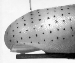
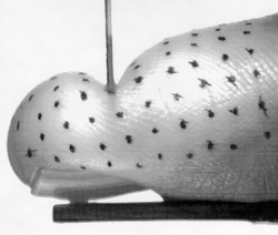
The experimental data was obtained by indenting human fingerpads with
several cylindrical and rectangular indentors and acquiring images of
the undeformed and deformed fingerpad using the videomicroscopy setup
(Roby, Dandekar, and Srinivasan, 1994; Roby and Srinivasan, 1995). Fine
markers were placed on the fingerpad and the skin surface deformation
was measured by tracking the displacements of the markers in the high
resolution video images. The same experiment was simulated using the finite
element models of the human fingertip and the displacements of corresponding
points were compared with the experimental data. The displacements predicted
by the multilayered 3D model matched the experimental data quite well
(Dandekar, 1995).
|
Click on the following links to read more in specific areas.
|
|
